Bones In Leg Diagram / horse skeleton diagram | Horse Anatomy Diagrams - The ... / Bones of the lower limb anatomy and physiology i.
byAdmin•
0
Bones In Leg Diagram / horse skeleton diagram | Horse Anatomy Diagrams - The ... / Bones of the lower limb anatomy and physiology i.. Anterior view with primary bones names. 2006 kia optima belt diagram. Top suggestions for human leg bones diagram. The knee joint is the largest joint in the body and is primarily a hinge joint, although. I had to reverse some numbers because the model i was working with has bird legs, but as soon as i figured that out this was immensely helpful.
3d viewer is not available. I had to reverse some numbers because the model i was working with has bird legs, but as soon as i figured that out this was immensely helpful. Leg bone wikipedia, femur bone diagram get rid of wiring diagram problem, amazon com poster foundry human bone anatomy illustration, knee common causes and symptoms stryker, bones of lower limb laminated anatomy chart. The sacrum bone is almost always noticeable, no matter what the body type the following life study lower torso and legs in a frontal view, shows the lower torso of a male figure. Most bones (particularly the long bones of the arms and legs — which make up the appendicular skeleton) have a hard outer shell known as cortical bone.
Leg Anatomy from www.fpnotebook.com Top suggestions for human leg bones diagram. When your muscles contract, they pull the bone they're. The foot bones shown in this diagram are the talus, navicular, cuneiform, cuboid, metatarsals and calcaneus. Explore more like human leg bones diagram. The human leg consists of 8 bones, 4 per leg. Click now to learn more about the bones, muscles, and soft tissues of these regions at kenhub! Posted on january 20, 2015 by admin. Learn more here you are seeing a 360° image instead.
The bones involved in it, however, are only the femur and the tibia, although the smaller bone of the leg, the fibula, is carried along in the movements of flexion, extension, and slight rotation that this joint permits.
Time to jump right into the biggest and strongest bones in the human body. Anterior view with primary bones names. Explore more like human leg bones diagram. The accompanying muscle diagram reveals the position of the muscles of the lower legs in this pose. The bones involved in it, however, are only the femur and the tibia, although the smaller bone of the leg, the fibula, is carried along in the movements of flexion, extension, and slight rotation that this joint permits. Leg bones labeled (page 1). The foot bones shown in this diagram are the talus, navicular, cuneiform, cuboid, metatarsals and calcaneus. Learn how to draw the femur, patella, tibia, and fibula in this lesson! The knee is a strong but flexible hinge joint. It allows the arm to come forward, out to the side. When your muscles contract, they pull the bone they're. For more details go to edit properties. Bones pain hand and arm bones diagram.
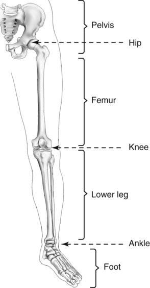
The human leg, in the general word sense, is the entire lower limb of the human body, including the foot, thigh and even the hip or gluteal region. Vector illustration with human skeleton scheme isolated on a white background. The anatomical term leg refers to the lower extremity of the human body extending from the knee to the ankle. Want to learn more about it? Time to jump right into the biggest and strongest bones in the human body.
Lower Limb and Pelvis | Radiology Key from radiologykey.com Most bones (particularly the long bones of the arms and legs — which make up the appendicular skeleton) have a hard outer shell known as cortical bone. The bones of your leg have roughened patches on their surfaces where muscles are attached. What does this suggest about mammals? What are the two bones in the lower arm called : Posted on january 20, 2015 by admin. The knee is a strong but flexible hinge joint. Human leg bones with telugu labels. Ankle and foot pain massage therapy connections.
He leg's main function in the human is for locomotion and support of the rest of the body.
Derivative of file:human leg bones labeled.svg which in turn is from file:human skeleton front en.svg. The accompanying muscle diagram reveals the position of the muscles of the lower legs in this pose. The knee is a strong but flexible hinge joint. The tibia (shin bone) is the medial bone of the leg and is larger than the fibula, with which it is paired (figure 3). Want to learn more about it? For more details go to edit properties. The bones of your leg have roughened patches on their surfaces where muscles are attached. Most bones (particularly the long bones of the arms and legs — which make up the appendicular skeleton) have a hard outer shell known as cortical bone. Learn more here you are seeing a 360° image instead. The bones of the leg are the femur, tibia, fibula and patella. Bones of the lower limb anatomy and physiology i. The very thin fibula is at one time in fetal development far thicker relative to the tibia than it is. Learn how to draw the femur, patella, tibia, and fibula in this lesson!
Your leg bones are very large and strong to help support the weight of your body. The tibia (shin bone) is the medial bone of the leg and is larger than the fibula, with which it is paired (figure 3). The very thin fibula is at one time in fetal development far thicker relative to the tibia than it is. The foot bones shown in this diagram are the talus, navicular, cuneiform, cuboid, metatarsals and calcaneus. Download a free preview or high quality adobe illustrator ai, eps, pdf and high resolution jpeg versions.
Human Leg Bone Structure - Human Anatomy Details from 2.bp.blogspot.com The human leg, in the general word sense, is the entire lower limb of the human body, including the foot, thigh and even the hip or gluteal region. Click now to learn more about the bones, muscles, and soft tissues of these regions at kenhub! B) that mammals are evolving to become more and more like one another. 2006 kia optima belt diagram. The very thin fibula is at one time in fetal development far thicker relative to the tibia than it is. It allows the arm to come forward, out to the side. I had to reverse some numbers because the model i was working with has bird legs, but as soon as i figured that out this was immensely helpful. Leg bones labeled (page 1).
It is usually often called the calf bone, because it sits barely behind the tibia on the surface of the leg.
When you stand or walk, all the weight of your upper body rests on them. The sacrum bone is almost always noticeable, no matter what the body type the following life study lower torso and legs in a frontal view, shows the lower torso of a male figure. Derivative of file:human leg bones labeled.svg which in turn is from file:human skeleton front en.svg. Upper leg bones diagram : The second largest bone in physique is the tibia, additionally known as the shinbone. Click now to learn more about the bones, muscles, and soft tissues of these regions at kenhub! Your leg bones are the longest and strongest bones in your body. Leg bone wikipedia, femur bone diagram get rid of wiring diagram problem, amazon com poster foundry human bone anatomy illustration, knee common causes and symptoms stryker, bones of lower limb laminated anatomy chart. What are the two bones in the lower arm called : Vector illustration with human skeleton scheme isolated on a white background. The femur, or thigh bone, is the largest, heaviest, and strongest bone in the human body. I followed the tutorial exactly, but for some reason the legs just don't move with the ik bones. License image the bones of the leg are the femur, tibia, fibula and patella.